Ray visualization and analysis of IOL optical performance
Heidelberg, 05 February 2020. Today, we are pleased to report the publication of our work on how to visualize light rays passing through IOLs, especially multifocal lenses.
Ray propagation imaging and optical quality evaluation of different intraocular lens models Hyeck Soo Son, Grzegorz Labuz, Ramin Khoramnia, Patrick Merz, Timur M. Yildirim, Gerd U. Auffarth. Published: February 4, 2020https://doi.org/10.1371/journal.pone.0228342
What we did.
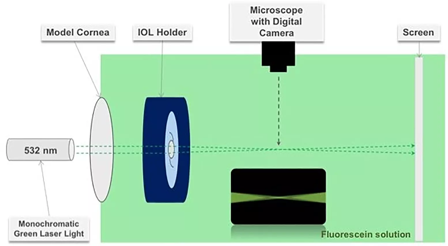
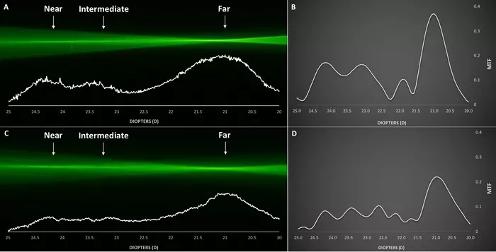
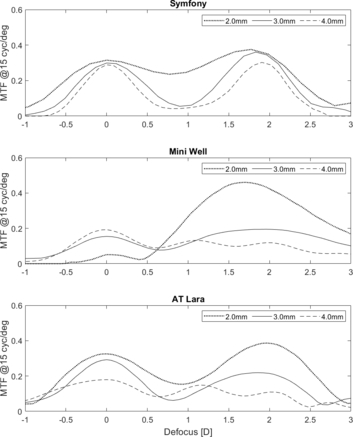
We assessed in the laboratory, four IOLs with different optical designs: a monofocal AcrySof IQ SN60WF [Alcon], a diffractive-refractive bifocal AcrySof IQ Restor SN6AD1 [Alcon], a diffractive trifocal AcrySof IQ PanOptix TFNT00 [Alcon], and a diffractive extended-depth-of-focus (EDOF) Symfony ZXR00 [Johnson&Johnson]. Our experimental set-up consisted of a water bath containing 0.01% fluorescein solution and a monochromatic green laser light source (532 nm) was used to visualize the propagation of light rays. We also evaluated the optical performance of these IOLs by measuring the modulation transfer function (MTF) values at a pupil sizes of 3.0 and 4.5 mm using the IOL-metrology device, OptiSpheric® IOL PRO II (Trioptics GmbH, Germany).
Our results are clear to see
Both the diffractive-refractive bifocal IOL and the EDOF IOL showed two defined foci for distance and near vision. In the diffractive trifocal IOL, three distinct foci for distance, intermediate, and near vision could be visualized, for example, the optical ray propagation and Through-Focus Response of the AcrySof IQ PanOptix TFNT00 at 3.0 mm (A, B) and 4.5 mm (C, D) pupil sizes.
What we concluded.
The ray propagation visualization technique allows a qualitative assessment and comparison of light energy distribution between different IOL models. The measured Through-Focus Response (TFR) quantitatively confirmed the evaluated ray propagation behaviour.
How to cite this paper
Article Source: Ray propagation imaging and optical quality evaluation of different intraocular lens models Son HS, Labuz G, Khoramnia R, Merz P, Yildirim TM, et al. (2020) Ray propagation imaging and optical quality evaluation of different intraocular lens models. PLOS ONE 15(2): e0228342. https://doi.org/10.1371/journal.pone.0228342
Go to PlosOne and the article citation can be downloaded in the following formats:
- RIS (compatible with EndNote, Reference Manager, ProCite, RefWorks)
- BibTex (compatible with BibDesk, LaTeX)
Acknowledgements
Funding & Support
We thank The Klaus Tschira Foundation, Heidelberg, Germany. The funding organization had no role in the design or conduct of this research.
end
 EDOF IOL research