
A new research article from the Apple Lab on IOL opacification after lamellar keratoplasty
Heidelberg 22 August 2017. The research group at the David J Apple International Laboratory for Ocular Pathology today published an online research paper through BMC Ophthalmology.
Hydrophilic intraocular lens opacification after posterior lamellar keratoplasty – a material analysis with special reference to optical quality assessment Bert C. Giers, Tamer Tandogan, Gerd U. Auffarth, Chul Y. Choi, Florian N. Auerbach, Saadettin Sel, Christian Mayer and Ramin Khoramnia. BMC Ophthalmology 2017 17:150
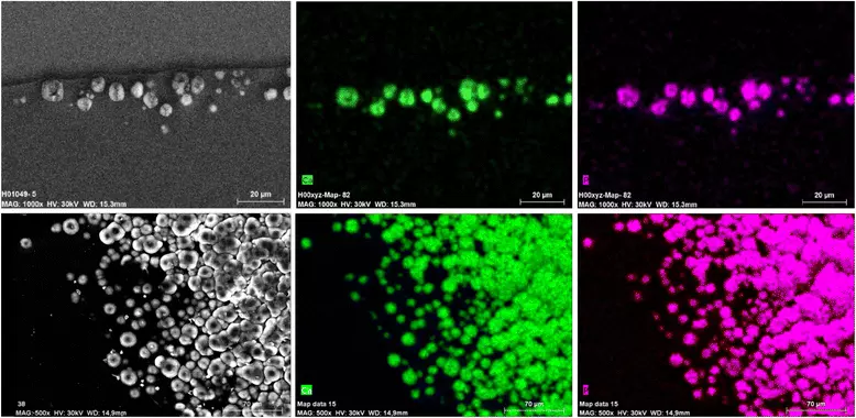
(The image above - introducing this article - is Exemplary Element Mapping showing calcium (green) and phosphorus (magenta) within the deposits in IOL #5.)
The Background
We conducted Laboratory analysis and optical quality assessment of explanted hydrophilic intraocular lenses (IOLs) with clinically significant opacification after posterior lamellar keratoplasty (DMEK and DSAEK).
Methods
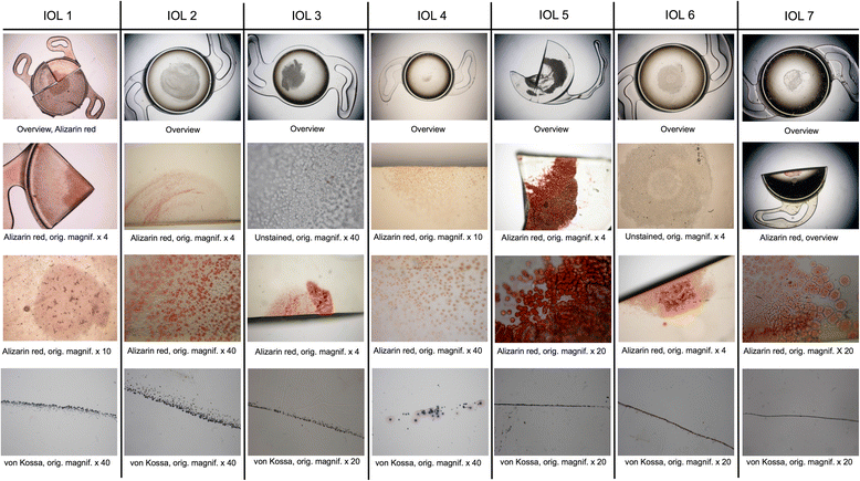
We analysed Thirteen opacified IOLs after posterior lamellar keratoplasty, 8 after Descemet stripping automated endothelial keratoplasty (DSAEK), 3 after Descemet membrane endothelial keratoplasty (DMEK) and 2 after both DSAEK and DMEK. Analyses included optical bench assessment for optical quality, light microscopy, scanning electron microscopy (SEM) and energy dispersive X-Ray spectroscopy (EDS).
Results
In all IOLs, the opacification was caused by a thin layer of Calcium phosphate that had accumulated underneath the anterior optical surface in the area spared by the pupil/anterior capsulorhexis. The calcifications lead to a significant deterioration of the modulation transfer function across all spatial frequencies of the affected IOLs.
IOLs 1–7. Light microscopy images representing an overview of the IOL optic and higher magnification images with Alizarin Red and von Kossa stainings.
Conclusions
Instilling an exogenous material such as air or gas into the anterior chamber increases the risk for opacification of hydrophilic IOLs irrespective of the manufacturer or the exact composition of the hydrophilic lens material. It is recommended to avoid the use of hydrophilic acrylic IOLs in patients with endothelial dystrophy that will likely require procedures involving the intracameral instillation of air or gas, such as DMEK or DS(A)EK.
About BMC …
BMC Ophthalmology is an open-access, peer-reviewed journal that considers articles on all aspects of the prevention, diagnosis and management of eye disorders, as well as related molecular genetics, pathophysiology, and epidemiology.
 (ARCHIVE) 2016 Heidelberg Helmholtz Symposium