Heidelberg, 29 March 2021: We are pleased to share the results of our latest study showing a morphological-functional correlation between the influence of an IOL’s localised calcification and straylight. What is especially novel about our study is the use two medical devices that are typically restricted to use in a diagnostic setting; we transfered their use to the laboratory. Our paper was published in the February edition of the American Journal of Ophthalmology.
American Journal of Ophthalmology.VOLUME 226, P108-116, JUNE 01, 2021 A Novel Approach for Assessing Visual Impairment Caused by Intraocular Lens Opacification: High-Resolution Optical Coherence Tomography Timur M. Yildirim, Grzegorz Łabuz, Maximilian Hammer, Hyeck-Soo Son, Sonja K. Schickhardt, Gerd U. Auffarth, Ramin Khoramnia DOI:https://doi.org/10.1016/j.ajo.2021.02.001
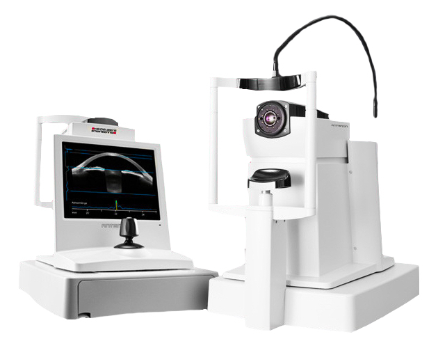
Top Photo: ANTERION®, Heidelberg Engineering GmbH, Heidelberg, Germany
Why did we study this?
Localized calcification occurs in pseudophakic eyes implanted with any hydrophilic IOL. It is relatively rare, and in previous studies we can link its development to the iatrogenic use of gases, which might damage or desiccate the IOL surface; and - apart from gases - other factors that change the intraocular microenvironment promoting deposition of Calcium compounds on or within the IOL polymer. Major clinical risk factors include secondary intraocular surgeries, especially Descemet membrane endothelial keratoplasty (DMEK) and pars plana vitrectomy (PPV).
DMEK has gained wide popularity and is increasingly performed at an earlier stage of the endothelial disease. Thus, we can expect to see some increase in patients with IOL localized calcification. It can be challenging, in such cases, solely from routine clinical examinations to assess the extent of visual impairment.
With the help of emerging high-resolution diagnostic imaging technologies, it is possible to visualize IOL opacification in-vitro without destroying the IOL. We used high-resolution optical coherence tomography to develop a way to predict intraocular straylight resulting from IOL localized opacification.
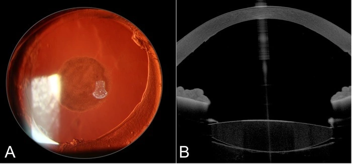
Photos A & B Clinical case of a patient with localized IOL opacification in the left eye following DMEK. A: Slitlamp photograph in retroillumination, B: Anterior segment optical coherence tomography image. Apart from the central IOL opacity, the attached DMEK lamella can be visualized at the cornea’s posterior side.
What did we do?
We obtained OCT images of forty-four IOLs that had been explanted intact between June 2018 and August 2020. In twenty-four cases, the IOL was removed due to a centrally localized opacification following either anterior or posterior secondary intraocular surgical procedures. The remaining twenty had been explanted because of IOL (sub)-luxation – and these clear (unopacified) IOLs acted as a control group.
In a novel in-vitro setup, we used a clinical anterior segment optical coherence tomography (AS-OCT) device, the ANTERION®, (Heidelberg Engineering, Heidelberg, Germany) - a device developed for clinal diagnostic use - to obtain high-resolution cross-section in-vitro images of all explants. Vertical B-scan-cross-section images were analyzed using ImageJ software. After applying a threshold, a standard rectangular Region of Interest (ROI) of 4 x 1 mm (as predefined in the software) was placed on the image in the lens optic center. The percentage of the area that was above the threshold within the ROI was determined. This numeric threshold area value was used as a metric for the amount of opacification in each IOL. Light scattering was quantified in all explants using an in-vitro setup with the second diagnostic device, the C-Quant straylight meter (Oculus GmbH, Wetzlar, Germany) which we have used quite routinely and is described in our previous papers.
We took the data and used a simple linear regression model was calculated based on the straylight and threshold area values.
What did we find?
We could visualize distinct morphological patterns in the twenty-four opacified IOLs using the Anterion device.
Image analysis resulted in a mean threshold area of 6.7 ±3.3 % and 2.0 ±0.8 % for the opacified IOL and the control group, respectively, P<0.05. The opacified lenses induced a mean straylight of 95.1 ±75.6 deg2/sr, whereas, in the control group, the straylight value was 5.0 ±3.4 deg2/sr, P<0.05.
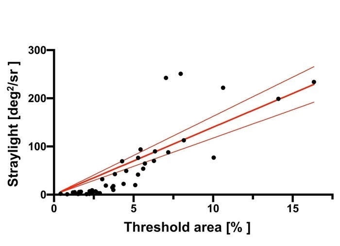 Chart: Threshold area (x-axis) and straylight value (y-axis) had a statistically significant correlation, with R2=0.80, P<0.001. The red lines show the fitted linear regression with 95% confidence intervals.
In a nutshell
- High-resolution optical coherence tomography visualized intraocular lens (IOL) opacities.
- The amount of IOL opacification correlated with the amount of induced straylight.
- Linear regression estimated the impact of IOL opacities on the visual quality.
Conclusions
This high-resolution OCT imaging technique can be used to visualize IOL opacities. The amount of opacification correlated well with the straylight induced by the lens. So we might expect that Anterior segment OCT imaging might be used in the future as a tool for predicting the extent of visual impairment and aid clinicians to quantify patients’ complaints.
Thanks
The David J. Apple International Laboratory for Ocular Pathology receives funding from the Klaus Tschira Stiftung, Heidelberg, Germany. T. Yildirim is supported by the Physician-Scientist Program of the Heidelberg University, Faculty of Medicine, Heidelberg. Funding organizations had no role in the design or conduct of this research.
We would like to thank Heidelberg Engineering GmbH, Heidelberg, for providing technical support during the course of this study. We appreciate the help of Mr. J. Mittenzwei, Mrs. U. Scheurich and Mr. W. Kreiter of the Department of Precision Engineering of the Heidelberg University for their help with constructing the IOL-holder for the in-vitro imaging.
Please find the full peer-reviewed AJO paper via this link:
https://www.ajo.com/article/S0002-9394(21)00063-5/fulltext
Cite this paper
Yildirim TM, Łabuz G, Hammer M, et al. A novel approach for assessing visual impairment caused by intraocular lens opacification: high-resolution optical coherence tomography: Straylight prediction in locally opacified IOLs. American Journal of Ophthalmology. 2021 Feb. DOI: 10.1016/j.ajo.2021.02.001.
end.
 IOL fixed to the capsulotomy!