Variation in IOL calcification in eyes with supplementary sulcus-supported lenses
Heidelberg, 27 July 2020. Contributed by Dr. Timur Yildirim. We are pleased to report the publication in the forthcoming September 2020 edition of the American Journal of Ophthalmology Case Reports, our work on IOL calcification in patients with supplementary IOLs.
Variation in intraocular lens calcification under different environmental conditions in eyes with supplementary sulcus-supported lenses Timur M. Yildirim, Ramin Khoramnia, Sonja K. Schickhardt, Donald J. Munro, Patrick R. Merz, Hyeck-Soo Son, Ingo Lieberwirth, Gerd U. Auffarth. https://doi.org/10.1016/j.ajoc.2020.100797
The report can be viewed and downloaded via, https://www.sciencedirect.com/science/article/pii/S245199362030133X
What we did
We teamed up with The Max Planck Institute for Polymer Research in Mainz, to analyze in a laboratory investigation, IOLs explanted from polypseudophakic eyes that had supplementary sulcus-supported IOLs. We analyzed both the supplementary lenses and capsular bag IOLs
All lenses were received at our Lab between January 2012 and March 2018. We studied each lens first with the light microscope (noting the morphology of opacification) next we used histological and electron microscopic techniques, and we evaluated the patients’ medical history
Analysis reveals Crystals
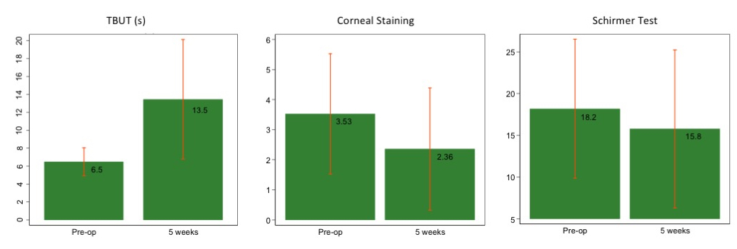
Eleven lenses were explanted due to IOL opacification from seven polypseudophakic eyes: In three cases, the supplementary lens calcified, in another three cases the opacification was in the capsular bag IOL (both lenses analyzed) and in one case both IOLs were opacified (we received only the supplementary IOL). For each case, we looked at the additional surgical procedures that had been performed since the IOL implantation, and these included pars plana vitrectomy or Descemet stripping endothelial keratoplasty. The most frequent comorbidity was diabetes mellitus.
Photographs of four cases of calcified sulcus-supported IOLs, the upper row shows 12.5-fold magnification overview images of the lenses. The second row - after staining of superficial deposits with Alizarin Red. The third row - sagittal cross-sections, after staining with von Kossa. We took these images in these three rows with a camera on a light microscope. The lowest row shows scanning electron microscopic images of the lenses’ cross-sections revealing different calcification patterns.
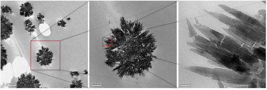
We used a technique new to this field: Transmission Electron Microscopy and electron diffraction pattern analysis, to investigate in detail the structure of the opacifying crystals.
For each opacified lens, we found a varying layer of calcium phosphate beneath the surface of the lens-optic. Crystal characterization revealed its composition to be Hydroxyapatite. The detailed morphology, down to 50nm, shows opacifying crystal structures clustered like flower petals.

Viewed at different magnifications - the microstructure of the hydroxyapatite crystal. These crystals were situated within the polymer material.
Conclusions.
This was a series of secondary calcification in lenses explanted from polypseudophakic eyes. In some cases, calcification occurred in the capsular bag lens, in other cases in the supplementary lens, or both. We could relate the severity of the morphological change to the comorbidities and the detail that after the lens implantation surgeries, there was subsequent ocular surgery.
Calcification of hydrophilic acrylic lenses is a continuing concern in IOL surgery. Lenses with this pathology can exhibit a reduced optical quality, and currently, the only treatment option is an IOL exchange (which itself can lead to further complications). In our series, the calcification occurred in eyes that had two IOLs, one primary capsular-fixated and one sulcus-supported lens. (To our knowledge, this is the most extensive series to-date of calcified supplementary IOLs.) Since both IOLs experienced the same intraocular environment, examining this series takes us a step closer to a fuller understanding of the origin and development of opacification. We expect that by continuing to study explanted calcified IOLs from polypseudophakic eyes and placing the results alongside the results of the ongoing modeling studies of lens opacification, we will get a much better understanding of IOL calcification.
Summary
- Intraocular lenses exhibited calcification in a series of polypseudophakic eyes.
- Pathology occurred in the capsular bag lens, the supplementary lens, or in both.
- Calcification was associated with different environmental factors.
- Depending on the factor, morphology, and pattern of the opacity differed.
- Using Transmission Electron Microscopy reveals the opacifying crystals’ ultrastructure.
How to cite this paper
- Timur M. Yildirim, Ramin Khoramnia, Sonja K. Schickhardt, Donald J. Munro, Patrick R. Merz, Hyeck-Soo Son, Ingo Lieberwirth, Gerd U. Auffarth, Variation in intraocular lens calcification under different environmental conditions in eyes with supplementary sulcus-supported lenses, American Journal of Ophthalmology Case Reports, Volume 19, 2020, 100797, ISSN 2451-9936
- https://doi.org/10.1016/j.ajoc.2020.100797.
- (http://www.sciencedirect.com/science/article/pii/S245199362030133X)
 Prizewinning work on Infra-red vision